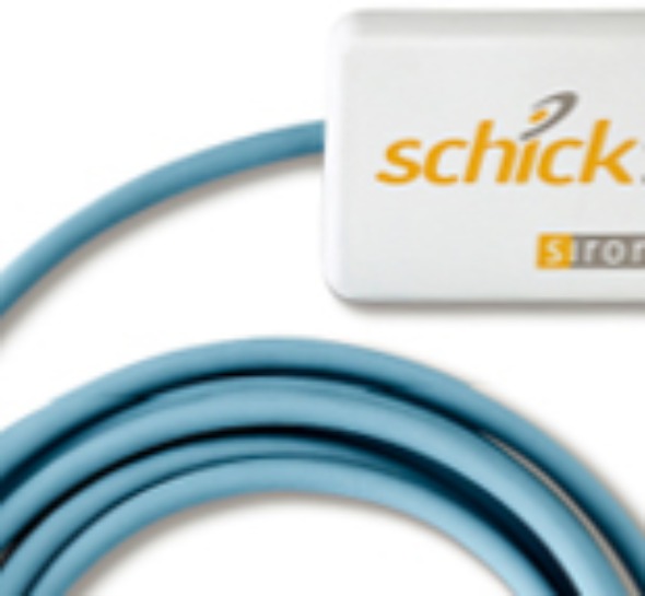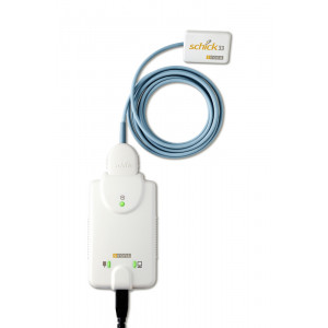

Conclusions: There was no sensor in the group that had the best physical design. For all sensor sizes, active imaging area was smaller compared with conventional films. The results of this study revealed that these details are not always available through the material provided by the manufacturers and are often not advertised. For most parameters tested, a lack of standardization exists in the industry. Results: There were variations among the physical design of different sensors. The sensors were evaluated for cable configuration, connectivity interface, presence of back-scattering radiation shield, plate thickness, active sensor area, and comparing the active imaging area to the outside casing and to conventional radiographic films.
#Schick 33 sensor size demension plus#
Methods: Sensors tested included: XDR (Cyber Medical Imaging, Los Angeles, CA, USA), RVG 6100 (Carestream Dental LLC, Atlanta, GA, USA), Platinum (DEXIS LLC., Hatfield, PA, USA), CDR Elite (Schick Technologies, Long Island City, NY, USA), ProSensor (Planmeca, Helsinki, Finland), EVA (ImageWorks, Elmsford, NY, USA), XIOS Plus (Sirona, Bensheim, Germany), and GXS-700 (Gendex Dental Systems, Hatfield, PA, USA). The purpose of this paper is to present the results of the evaluation of the physical design of eight CMOS digital intraoral sensors.

Objective: Digital technologies provide clinically acceptable results comparable to traditional films while having other advantages such as the ability to store and manipulate images, immediate evaluation of the image diagnostic quality, possible reduction in patient radiation exposure, and so on.


 0 kommentar(er)
